Radiation has become a cornerstone in medical imaging, enabling doctors to peer inside the body and make critical diagnoses without invasive procedures. However, as with any powerful tool, radiation carries risks, and one of the most overlooked yet significant dangers is scattered radiation. Scattered radiation, also called secondary radiation, poses exposure risks to both patients and healthcare providers. Understanding its effects and implementing safety protocols is essential to mitigate these risks in medical imaging environments.
What Is Scattered Radiation?
Scattered radiation is a form of radiation that deviates from its intended path as it interacts with objects, such as a patient’s body. In medical imaging, primary radiation beams target specific areas to capture diagnostic images. However, when this radiation encounters tissues, organs, and bones, a portion of it can be deflected, resulting in scattered radiation that travels unpredictably in different directions. Unlike the primary beam, which is focused and controlled, scattered radiation disperses within the imaging room and potentially exposes surrounding individuals to unnecessary radiation.
Types of Scattered Radiation in Medical Imaging
The interaction of X-rays with human tissue during medical imaging procedures produces two main types of scattered radiation:
- Compton Scattering: Occurs when an X-ray photon collides with a loosely bound electron in an atom, causing the electron to be ejected and the X-ray photon to scatter in another direction. This is the most common type of scattering in diagnostic imaging, especially at higher energies.
- Coherent (Rayleigh) Scattering: Involves low-energy photons that interact with the entire atom without ionizing it, resulting in the photon scattering without a significant energy loss. Although less common, this form of scattering also contributes to exposure risks in specific imaging procedures.
The Impact of Scattered Radiation on Exposure Risks
An effect of scattered radiation is to increase exposure risk, a concern that becomes particularly significant in high-radiation settings, such as radiography, fluoroscopy, and CT scans. Scattered radiation does not contribute to the formation of the medical image; instead, it disperses, exposing not only the patient but also the healthcare professionals present in the room. Over time, repeated exposure can lead to cumulative radiation doses, which are associated with increased health risks.
Exposure Risks for Patients
For patients, scattered radiation can lead to unintended radiation doses beyond the targeted area. Although these doses are generally lower than the primary radiation dose, they still add to the overall radiation exposure, which may have cumulative effects, especially in patients requiring repeated imaging. Increased radiation exposure over time has been associated with a heightened risk of radiation-induced conditions, such as skin erythema, tissue damage, and, in extreme cases, a higher lifetime risk of cancer.
Exposure Risks for Medical Personnel
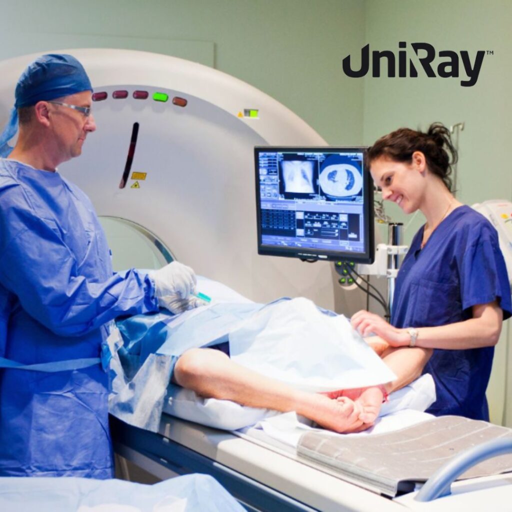
Medical personnel, particularly radiologists, technicians, and interventionalists, are at higher risk of exposure due to the cumulative nature of radiation exposure over their careers. Scatter from frequent procedures increases their likelihood of receiving a significant cumulative dose. Prolonged exposure to scattered radiation can lead to occupational health issues, including cataracts, skin damage, and increased cancer risk. Thus, understanding and minimizing exposure to scattered radiation is essential to protect healthcare providers’ long-term health.
How Scattered Radiation Affects Image Quality
Beyond health risks, scattered radiation impacts the quality of diagnostic images. As scattered radiation enters the detector from unintended angles, it creates noise in the image, reducing contrast and clarity. This effect can make it harder to distinguish between different tissues, potentially leading to missed diagnoses or the need for repeat imaging, both of which elevate the patient’s overall exposure. Techniques to minimize scattered radiation, such as collimation and grid use, play a critical role in maintaining image quality and diagnostic accuracy.
Techniques to Reduce the Impact of Scattered Radiation on Image Quality
To enhance image clarity and reduce the negative effects of scattered radiation, radiologic technologists employ various methods:
- Use of Anti-Scatter Grids: Grids are positioned between the patient and the detector to filter out scattered radiation, allowing only the primary beam to reach the detector. This significantly enhances image contrast, though it may increase the primary radiation dose required.
- Collimation: By narrowing the X-ray beam to the area of interest, collimation reduces the amount of tissue exposed to radiation, consequently decreasing scattered radiation. This technique minimizes radiation exposure while enhancing image quality.
- Air Gaps: Placing a distance between the patient and the detector creates an “air gap,” which allows some scattered radiation to diverge away from the detector. This reduces scatter reaching the imaging detector and improves image contrast, though it is more commonly used in mammography.
Regulatory Guidelines and Safety Standards for Managing Scattered Radiation
Given the potential health impacts of scattered radiation, regulatory agencies have established safety standards to protect patients and staff. Organizations such as the International Commission on Radiological Protection (ICRP), the National Council on Radiation Protection and Measurements (NCRP), and the American College of Radiology (ACR) provide guidelines to optimize radiation use and minimize unnecessary exposure.
Key Recommendations for Scattered Radiation Safety
- ALARA Principle: The ALARA (As Low As Reasonably Achievable) principle is foundational to radiation safety, encouraging all healthcare settings to minimize exposure to the lowest level possible without compromising diagnostic efficacy.
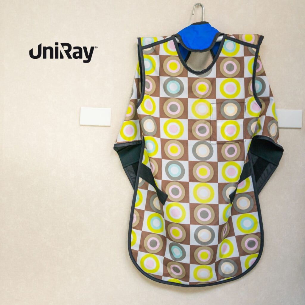
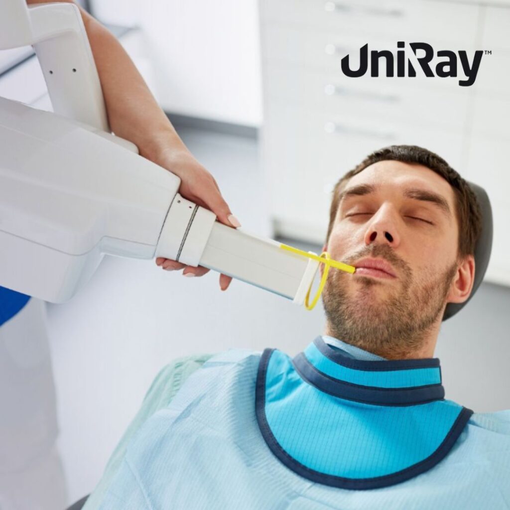
- Personal Protective Equipment (PPE): Lead apron, thyroid shield, lead gloves, and other radiation protection gear are essential for medical personnel exposed to scattered radiation during procedures.
- Radiation Monitoring: Personal dosimeters measure cumulative radiation exposure to ensure it remains within safe limits. Regular monitoring helps identify and address any breaches in safety protocols.
- Shielding Barriers: Fixed or mobile lead barriers provide an additional layer of protection for medical staff during procedures, particularly in fluoroscopy and interventional radiology.
Minimizing Exposure to Scattered Radiation: Best Practices
Given that an effect of scattered radiation is to increase exposure risk, medical facilities have implemented best practices for minimizing exposure. Here are some standard methods used in clinical settings:
- Proper Patient Positioning and Beam Alignment: Ensuring the patient and the X-ray beam are aligned reduces the amount of radiation required, indirectly minimizing scatter. Accurate positioning also decreases the need for repeat exposures.
- Optimized Imaging Parameters: Adjusting technical factors, such as the tube current (mA) and exposure time, helps to use the minimum radiation dose necessary while achieving quality imaging. Lower radiation output correlates with reduced scatter.
- Use of Pulsed Fluoroscopy: In fluoroscopic procedures, pulsed fluoroscopy rather than continuous X-ray use can reduce radiation exposure for both the patient and staff. This technique emits radiation in intervals, minimizing scatter without compromising image quality.
- Limit Time in the Room: For staff members, limiting the time spent in a radiation field reduces cumulative exposure. Time, distance, and shielding—often called the “three pillars of radiation safety”—are essential concepts for managing exposure in healthcare settings.
- Educational Programs: Continuous education and training for healthcare providers on radiation safety techniques ensure that best practices are consistently implemented and followed.
Technological Advancements in Reducing Scattered Radiation
As technology advances, so do methods for reducing the impact of scattered radiation. Digital imaging systems now feature software that can detect and correct for scatter, enhancing image quality without additional hardware. Innovations such as digital subtraction angiography (DSA) can reduce scattered radiation by isolating areas of interest, further minimizing exposure. Additionally, low-dose CT imaging has become increasingly popular, allowing for high-quality imaging at a fraction of traditional radiation doses.
The Role of the Radiologic Technologist in Managing Scattered Radiation
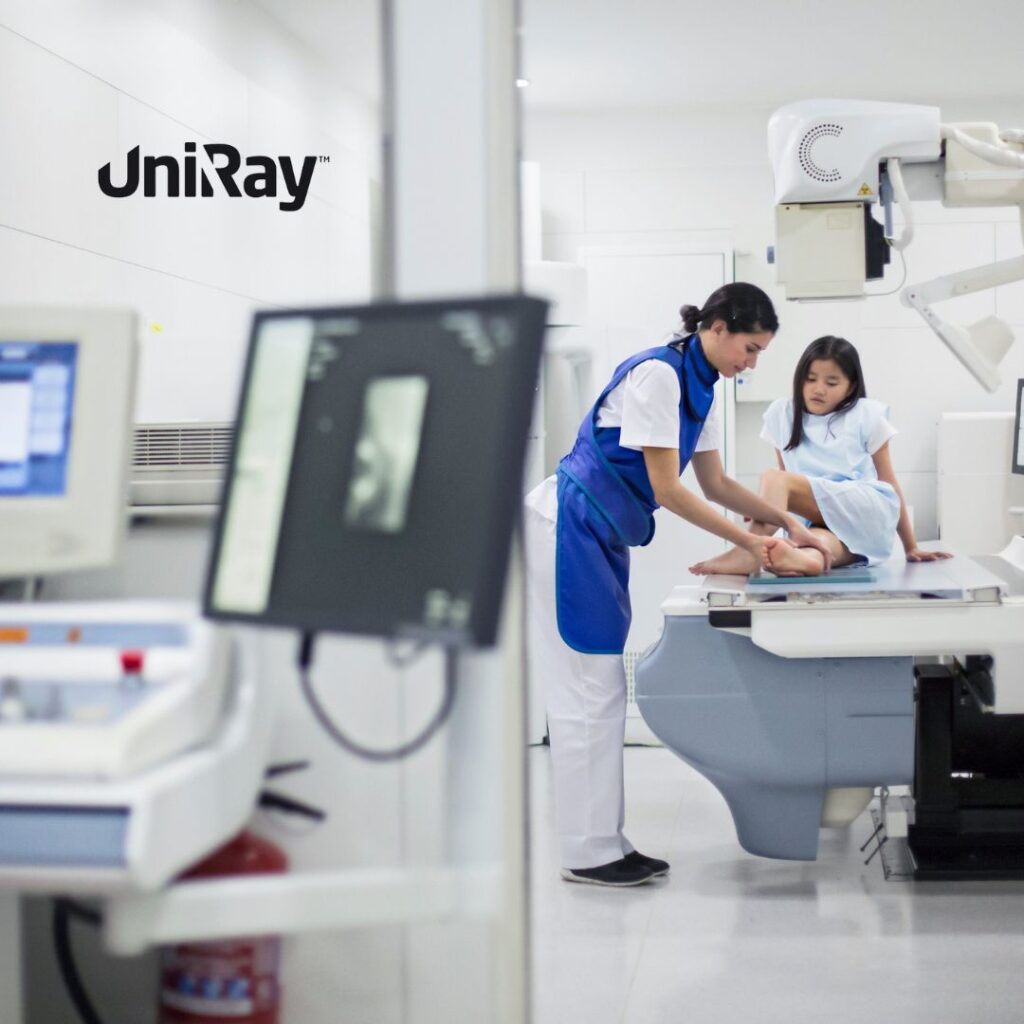
Radiologic technologists play a critical role in minimizing scattered radiation exposure. Their responsibilities include positioning patients, selecting appropriate equipment settings, and implementing radiation safety protocols. Through training, radiologic technologists become experts in both imaging and radiation protection, bridging the gap between diagnostic effectiveness and patient/staff safety. By following protocols, technologists can ensure that each procedure minimizes exposure while maintaining high diagnostic standards.
Conclusion
The widespread use of medical imaging has revolutionized healthcare, but it also brings responsibility. An effect of scattered radiation is to increase exposure risk, posing potential health hazards for both patients and medical professionals. Through awareness, technological advancements, and adherence to safety protocols, healthcare facilities can mitigate these risks and continue to use medical imaging as a powerful diagnostic tool. By implementing best practices and continually educating healthcare workers on safety standards, we can ensure that radiation’s benefits outweigh its risks, fostering a safer environment in medical imaging.


