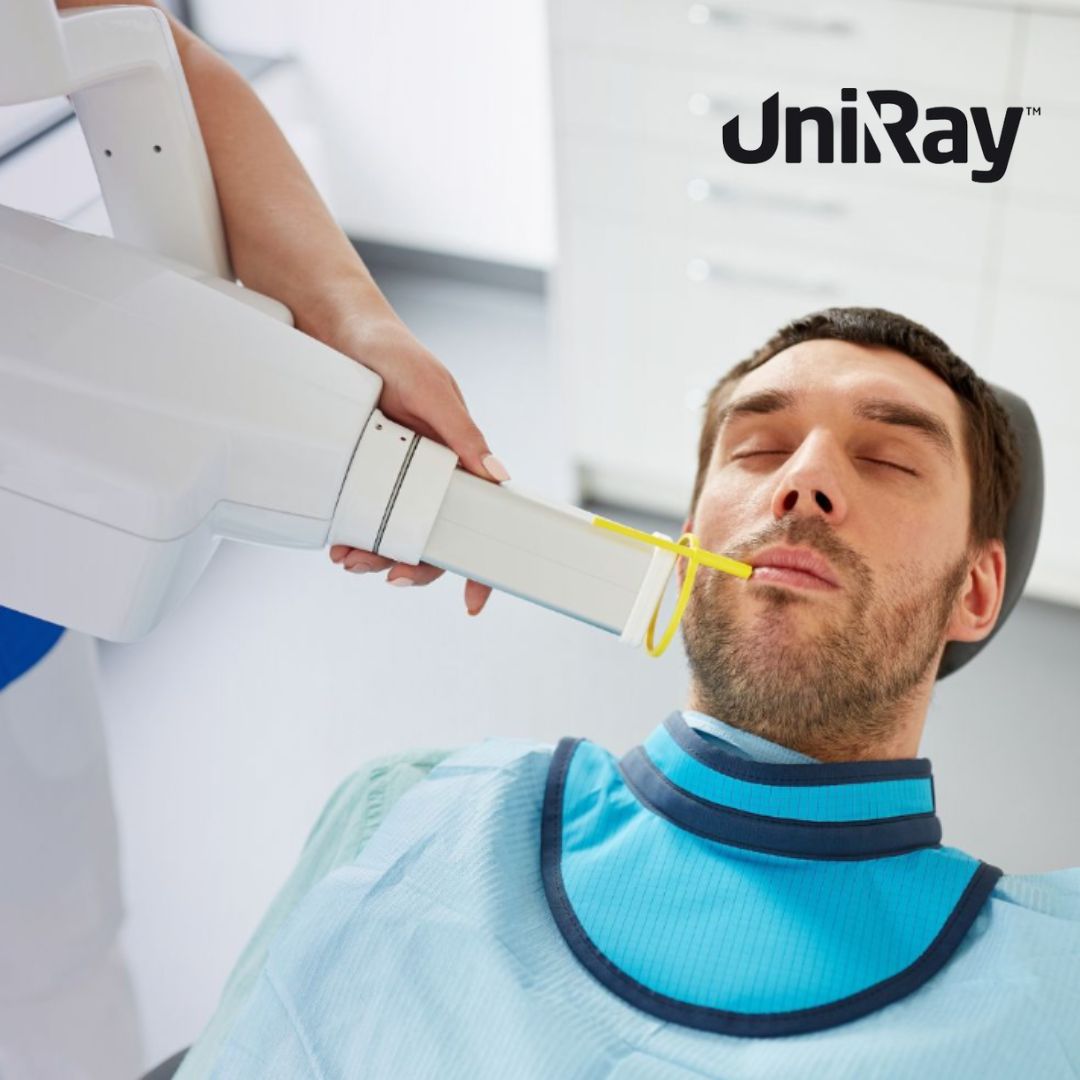
Dental radiographs, commonly known as X-rays, are an essential tool in modern dentistry. They allow dentists to diagnose problems that are not visible during a regular oral examination, such as decay between teeth, bone loss, or impacted teeth. However, like all forms of radiation, dental X-rays pose a potential risk if not used correctly. To minimize these risks and ensure patient safety, it is crucial to follow established dental radiation guidelines and adhere to best practices for dental X-ray recommendations. This blog will explore the importance of radiation safety in dental care, outline essential guidelines, and provide key recommendations for the use of X-rays in dental settings.
The Importance of Dental Radiation Guidelines
Radiation exposure, even in small doses, can have cumulative effects over time. While the risk associated with dental X-rays is relatively low compared to other medical imaging modalities, it is still essential to minimize unnecessary exposure. Dental radiation guidelines have been developed to help protect both patients and dental professionals from excessive radiation exposure. These guidelines are grounded in the principles of ALARA (As Low As Reasonably Achievable), which emphasizes minimizing exposure while achieving the necessary diagnostic outcomes.
Dental professionals are required to take steps to ensure that the benefits of taking X-rays outweigh the risks, particularly for vulnerable populations such as children and pregnant women. By adhering to established guidelines, dental clinics can maintain a balance between patient safety and effective diagnostic practices.
Overview of Dental Radiation Guidelines
- Justification of X-Ray Use: According to dental radiation guidelines, X-rays should only be taken when necessary. Dentists must weigh the potential benefits of the radiograph against the risk of radiation exposure. Routine or unnecessary X-rays should be avoided, and decisions should be made on a case-by-case basis, considering the patient’s medical and dental history.
- Limiting Exposure: One of the core principles of dental radiation guidelines is to minimize patient exposure to radiation. This can be achieved through proper equipment maintenance, use of protective devices like dental lead apron and thyroid shield, and using the lowest possible radiation dose that will still provide diagnostic-quality images.
- Use of Digital Imaging: Modern dental practices are encouraged to use digital radiography, which exposes patients to significantly lower radiation levels compared to traditional film-based X-rays. Digital X-rays also offer the advantage of instant image availability and can be easily stored and shared electronically.
- Shielding and Protection: Dental radiation guidelines recommend the use of lead aprons and thyroid collar to shield patients from scatter radiation. The thyroid gland, in particular, is highly sensitive to radiation, and protecting it is essential in reducing radiation-related risks.
- Education and Training: Dental professionals must receive appropriate training on the safe use of radiographic equipment. This includes understanding the principles of radiation physics, proper positioning of the X-ray machine, and techniques to reduce exposure. Continuous education and staying updated on the latest dental radiation guidelines are crucial to maintaining a safe environment.
 Dental X-Ray Recommendations for Different Patient Groups
X-rays play a critical role in diagnosing dental conditions, but their use should be based on the specific needs of each patient. Here are the dental X-ray recommendations for different patient groups:
- Children: Children are more sensitive to radiation than adults due to their developing tissues and organs. As a result, dental X-ray recommendations for pediatric patients emphasize minimizing radiation exposure. Dentists should only take X-rays when absolutely necessary, and they should use child-sized settings on the X-ray machine. Protective shielding, such as lead aprons and thyroid collars, should always be used for children. Furthermore, digital radiography is especially recommended for pediatric patients due to its lower radiation doses.
- Pregnant Women: For pregnant patients, the general rule is to avoid dental X-rays unless they are urgently needed to treat an infection or other serious dental issue. If an X-ray is necessary, precautions must be taken to shield the abdomen and thyroid, protecting both the mother and the developing fetus. The American Dental Association (ADA) suggests that routine X-rays can often be postponed until after pregnancy, but urgent dental care, including the use of X-rays when essential, should not be delayed.
- Adults with High-Risk Dental Issues: Adults with a history of extensive dental work, such as crowns, bridges, or implants, may require more frequent X-rays to monitor the condition of these restorations. Patients with periodontal disease or other chronic dental conditions may also need more frequent radiographs to track the progression of their condition and to ensure proper treatment.
- Adults Without Symptoms: For adults without dental symptoms or a history of extensive dental work, the dental X-ray recommendations suggest taking X-rays every 18 to 36 months, depending on the patient’s risk factors for dental diseases. Routine X-rays are not necessary if the patient is in good oral health and has no signs of decay or infection.
Types of Dental X-Rays and Their Applications
Understanding the different types of dental X-rays is essential for following dental radiation guidelines. Each type of radiograph serves a specific diagnostic purpose and should be used accordingly.
- Bitewing X-Rays: These X-rays show the upper and lower teeth in a specific section of the mouth. They are typically used to detect decay between teeth and to check for bone loss caused by gum disease. Bitewing X-rays are one of the most common types of dental radiographs and are often taken during routine dental checkups.
- Periapical X-Rays: These images show the entire tooth, from the crown to the root, and are useful for detecting issues with the tooth’s root or surrounding bone structure. Periapical X-rays are often used when diagnosing an abscess, fracture, or impacted tooth.
- Panoramic X-Rays: A panoramic X-ray provides a broad view of the entire mouth, including the teeth, jaws, and surrounding structures. This type of radiograph is particularly useful for identifying impacted teeth, detecting tumors, or assessing the overall condition of the jawbone. Panoramic X-rays are commonly used for patients undergoing orthodontic treatment or those requiring extensive dental surgery.
- Cone Beam Computed Tomography (CBCT): CBCT is a more advanced form of imaging that provides 3D images of the teeth, bones, and soft tissues. It is often used for planning dental implants, root canals, or complex oral surgeries. While CBCT exposes patients to more radiation than traditional X-rays, it provides invaluable information for intricate procedures.
Minimizing Radiation in Dental X-Rays
As previously mentioned, following the principle of ALARA is essential in adhering to dental radiation guidelines. Below are some additional recommendations for minimizing radiation exposure during dental X-rays:
- Use of Proper Equipment: Dental offices should regularly inspect and maintain their X-ray equipment to ensure that it is functioning properly and producing the lowest possible radiation dose. Regular calibration of the X-ray machine is necessary to avoid excess radiation exposure.
- Collimation and Filtration: Collimators are devices that restrict the size and shape of the X-ray beam, focusing it only on the area of interest. Proper collimation reduces scatter radiation and minimizes exposure to surrounding tissues. Additionally, the use of filters can remove low-energy X-rays that do not contribute to image formation, further reducing patient exposure.
- Proper Patient Positioning: Dental professionals must ensure that the patient is positioned correctly before taking an X-ray. Poor positioning can result in the need for retakes, leading to unnecessary exposure to radiation. Ensuring accurate positioning from the start helps avoid this issue.
- Use of Faster Image Receptors: Faster image receptors, such as digital sensors, require less radiation to produce clear images compared to traditional film. Digital imaging is now considered the gold standard in dental radiography due to its ability to reduce radiation doses while maintaining high-quality diagnostic images.
- Limiting Retakes: Dentists should strive to capture accurate images on the first attempt to avoid retaking X-rays. This requires proper training in radiographic techniques and attention to detail during the imaging process.
Conclusion
Dental X-rays are an invaluable tool in diagnosing and treating dental conditions, but they must be used responsibly to minimize the risks associated with radiation exposure. Adhering to dental radiation guidelines is essential in ensuring patient and staff safety. By following established recommendations, such as limiting exposure, using protective devices, and employing digital imaging, dental professionals can maintain high standards of care while minimizing radiation-related risks. Additionally, tailoring dental X-ray recommendations to the specific needs of individual patients helps achieve a balance between diagnostic efficacy and safety. Ultimately, by prioritizing radiation safety, dental practices can ensure that patients receive the benefits of radiography without unnecessary risks.