Dental Radiograph Guidelines: Ensuring Safety and Accuracy in Diagnostic Imaging
Dental radiographs, also known as dental X-rays, are critical tools in modern dentistry, enabling the early detection and diagnosis of a wide range of oral health issues that may not be visible to the naked eye. From cavities to bone loss and impacted teeth, these images allow dentists to create effective treatment plans. However, because dental X-rays expose patients to low levels of radiation, adhering to established guidelines is essential for ensuring both safety and accuracy. This comprehensive guide explores the importance of dental radiograph guidelines, proper safety procedures, and best practices for using dental radiographs effectively.
Understanding the Importance of Dental Radiographs
Dental X-rays are essential for providing dentists with an inside look at areas that cannot be easily viewed during routine examinations. They reveal cavities between teeth, infections at the root of the tooth, abscesses, tumors, impacted teeth, and even bone loss due to periodontal disease. Radiographs are also used to monitor the development of children’s teeth and track progress in orthodontic treatments.
Despite their diagnostic benefits, dental X-rays involve exposure to small amounts of radiation. Therefore, following dental radiograph guidelines helps balance the necessity of obtaining these images with the responsibility of limiting radiation exposure to both patients and dental professionals.
Types of Dental Radiographs
 Dental radiographs can be categorized into several types, each designed to capture specific details:
- Bitewing Radiographs: These show the upper and lower teeth in one section and are useful for detecting cavities between teeth.
- Periapical Radiographs: These capture the entire tooth, from the crown to the root, and are ideal for identifying tooth or bone abnormalities.
- Panoramic Radiographs: These provide a broad view of the entire mouth, including the jaws, teeth, and surrounding structures. They are often used for orthodontic treatment and in preparation for implants or extractions.
- Occlusal Radiographs: These are used to view the floor or roof of the mouth and are commonly used to find extra teeth, impacted teeth, or jaw fractures.
Each type of radiograph serves a distinct purpose, and the choice of which to use depends on the patient’s specific needs and the dentist’s clinical judgment.
Dental X-ray Safety and Radiation Exposure
 While dental X-rays are necessary, safety is a priority. The radiation exposure from dental X-rays is minimal compared to other types of medical imaging, but it still requires careful management. Radiographic equipment and techniques have improved significantly, reducing radiation levels even further. Digital X-rays, for example, use 70-90% less radiation than traditional film X-rays.
Key safety protocols to reduce radiation exposure include:
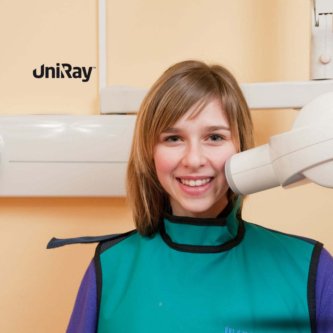
- Using Protective Gear: Patients should always wear lead aprons and thyroid collars to protect sensitive areas from unnecessary exposure.
- Minimizing Unnecessary X-rays: Dentists should follow guidelines to determine the necessity of X-rays based on the patient’s age, health history, and risk factors for dental disease.
- Maintaining Proper Equipment: Regular maintenance and calibration of radiographic equipment are essential to ensure images are captured efficiently with the lowest possible dose of radiation.
- Using the ALARA Principle: “As Low As Reasonably Achievable” is the guiding principle to minimize radiation exposure during radiographic procedures.
Dental Radiograph Guidelines for Special Populations
Certain groups require additional care when it comes to dental radiographs:
- Children: Pediatric patients are more sensitive to radiation, so the frequency and type of X-rays should be limited. Dentists must assess each child’s risk for dental disease before recommending X-rays.
- Pregnant Patients: Dental X-rays should be avoided during pregnancy unless absolutely necessary. If X-rays are essential, lead aprons and thyroid collars should be used to shield the abdomen and thyroid.
- High-Risk Patients: Individuals with a history of radiation therapy, autoimmune conditions, or other health concerns may require adjustments to their radiographic treatment plan to minimize risks.
How Often Should Dental X-rays Be Taken?
The frequency of dental X-rays depends on the patient’s individual oral health needs. The American Dental Association (ADA) provides general guidelines, but the dentist’s clinical judgment should ultimately determine the timing:
- Adults with good oral health: X-rays may only be needed every 2-3 years.
- Adults with a history of dental problems: More frequent X-rays may be necessary, possibly every 6-12 months.
- Children and adolescents: X-rays may be required more frequently as their teeth and jaws develop and shift. For instance, bitewing X-rays may be needed every 6-12 months if the child is prone to cavities.
Proper Dental Radiograph Procedures
For safe and effective radiographic imaging, dental professionals must follow proper procedures:
- Pre-Radiographic Assessment: The dentist should evaluate the patient’s dental and medical history and perform a clinical exam before deciding to take X-rays.
- Proper Positioning: Correct positioning of the patient and the X-ray machine ensures that clear images are obtained with minimal retakes.
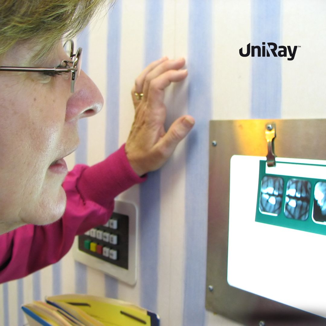
- Image Evaluation: After capturing the radiograph, it’s essential to thoroughly evaluate the image for clarity, accuracy, and diagnostic value. If the image quality is poor, a retake may be necessary, but only after careful consideration.
Advances in Digital Dental Radiography
 Digital radiography has become the standard in most dental practices due to its numerous advantages over traditional film X-rays. These benefits include:
- Lower Radiation Exposure: As mentioned earlier, digital X-rays reduce radiation exposure by up to 90%.
- Faster Image Capture: Digital radiographs provide instant results, reducing the time patients spend in the chair and allowing for immediate diagnosis.
- Enhanced Image Quality: Digital images can be enlarged, sharpened, and contrasted to enhance diagnostic accuracy.
- Environmentally Friendly: Digital radiography eliminates the need for film processing chemicals, making it more eco-friendly.
Best Practices for Dental Radiograph Storage and Management
Dental radiographs are important medical records, and proper storage and management are crucial. Digital radiographs can be stored electronically, making it easier to access and share them with other healthcare providers when necessary. HIPAA (Health Insurance Portability and Accountability Act) guidelines must be followed to protect the privacy of patients’ radiographic data. Regular backups of digital records are also necessary to prevent data loss.
Legal and Ethical Considerations in Dental Radiographs
Dentists have a legal and ethical obligation to limit radiation exposure and use radiographs judiciously. Overuse of dental X-rays can expose patients to unnecessary risks and lead to legal repercussions. Informed consent is another critical component—patients should always be informed of the need for X-rays, the benefits, and the risks involved. Proper documentation of radiographic findings and their justification is essential for maintaining patient trust and meeting legal standards.
Common Mistakes to Avoid in Dental Radiography
There are several common mistakes that can occur during dental radiography:
- Over-Radiographing: Taking X-rays too frequently or without clinical justification can unnecessarily expose patients to radiation.
- Improper Shielding: Failing to provide adequate protective gear, like lead aprons and thyroid collars, puts patients at risk.
- Poor Positioning: Incorrect patient positioning can result in unclear images, leading to retakes and increased radiation exposure.
- Failure to Follow Up: Neglecting to properly analyze radiographs or recommend appropriate treatment based on the findings can delay necessary care.
Conclusion: Ensuring Safe and Effective Dental Radiographs
Dental radiographs are an invaluable tool for diagnosing and treating a wide range of oral health issues. By following established dental radiation guidelines, including the use of protective measures and adherence to best practices, dental professionals can ensure the safety and well-being of their patients while benefiting from the diagnostic power of radiographs. With advancements in digital radiography, patients are exposed to less radiation than ever before, but it remains critical to use these tools judiciously. Proper training, patient communication, and a commitment to safety should be at the heart of every dental radiographic procedure.