Dental Radiation Safety & Protocol for Dental Professionals: A Comprehensive Guide
In the field of dentistry, the use of radiographs is essential for diagnosis and treatment planning. However, the risks associated with radiation exposure must not be overlooked. Dental Radiation safety & protocol for dental professionals is paramount to ensure the well-being of both patients and dental staff. This detailed guide covers key safety measures, protocols, and the latest guidelines that dental professionals should follow to minimize exposure risks while maintaining high diagnostic standards.
Understanding Radiation in Dental Practice

Dentists use X-rays (radiographs) for a variety of diagnostic purposes, such as detecting cavities, bone loss, impacted teeth, and other oral conditions. While the benefits of dental radiography are undeniable, the potential health risks posed by ionizing radiation should be addressed through proper safety protocols. Long-term exposure to radiation, even in small doses, can result in harmful effects like tissue damage and increased cancer risk.
That’s where dental radiation safety for professionals comes into play. Proper training, equipment, and protocols can significantly reduce radiation exposure and protect both patients and healthcare workers.
The Importance of Radiation Safety in Dental Offices
Dental professionals, including dentists, dental assistants, and hygienists, are exposed to radiation regularly. Though the doses are typically low, cumulative exposure over a long period can lead to adverse effects. By adhering to dental radiation protection in radiology guidelines, these risks can be mitigated.
It is crucial for dental clinics to follow strict radiation safety procedures for dental offices to safeguard everyone involved in the dental treatment process. These procedures involve everything from the type of radiographic equipment used to protective gear, patient positioning, and regulatory compliance.
Key Components of a Radiation Safety Program
1. Training and Education: All dental professionals should undergo regular training on radiation safety and radiographic techniques. This includes staying updated on the latest equipment and procedures that reduce exposure levels.
2. Equipment Maintenance and Upgrades: Dental radiography equipment must be regularly inspected and calibrated to ensure it operates at optimal safety levels. Modern digital X-ray machines expose patients and dental professionals to much less radiation compared to older film-based systems.

3. Radiation Monitoring Devices: Dental offices should use dosimeters to monitor radiation exposure for staff. These devices track cumulative radiation exposure, allowing dental professionals to ensure they are within safe limits.
4. Adherence to Dental Radiograph Guidelines: Strict adherence to dental radiograph guidelines ensures that radiographs are only taken when absolutely necessary. Overprescribing radiographs not only exposes patients and staff to unnecessary radiation but also increases healthcare costs.
Dental Lead Apron Requirements: An Essential Safety Measure

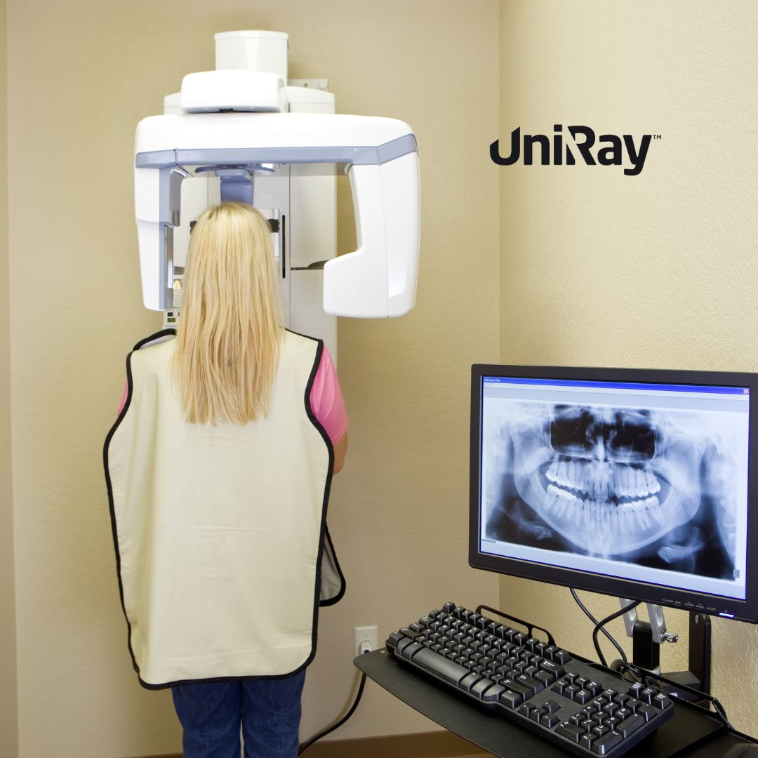
One of the most effective tools in radiation protection is the use of a dental apron. Dental lead apron requirements are an integral part of every dental clinic’s radiation safety protocol. Dental aprons shield vital organs such as the thyroid, chest, and abdomen from scattered radiation during dental radiographic procedures.
Key points regarding dental lead-free apron use include:
- Material and Thickness: Dental lead-free aprons are typically made from non-lead, lead-equivalent materials and must meet specific thickness standards to effectively block radiation. The recommended thickness for lead-free aprons in dental radiography is generally 0.25 mm of lead equivalence.
- Thyroid Collar: The thyroid is especially sensitive to radiation, making a thyroid collar an important addition to the dental apron. Patients should wear a thyroid collar whenever intraoral radiographs are taken.
- Proper Use and Storage: Dental professionals should ensure that lead-free aprons are properly placed on the patient, covering the necessary areas. These aprons should be stored flat or hung properly to prevent cracks, which could reduce their effectiveness.
- When to Use Lead-Free Aprons: While the use of digital radiography has reduced radiation exposure levels, most state and local regulations still mandate the use of lead-free aprons for patients during X-rays. Additionally, dental personnel should use aprons when in close proximity to the radiation source.
Radiation Protection in Dental Radiology: ALARA Principle
The guiding principle for radiation protection in dental radiology is the ALARA principle, which stands for “As Low As Reasonably Achievable.” This means that dental professionals should always aim to minimize radiation exposure to the lowest level possible while still achieving the desired diagnostic outcomes.
Key practices include:
- Minimizing Retakes: One of the most effective ways to reduce radiation exposure is to avoid unnecessary retakes of X-rays. Proper positioning and technique should be used to get the image right the first time.
- Use of Collimators and Filtration: Dental X-ray machines should be equipped with collimators that narrow the beam to the area of interest, reducing the radiation dose to the patient. Filtration systems can further block out unnecessary lower-energy radiation.
- Digital Radiography: Switching to digital X-rays instead of traditional film-based systems can reduce radiation exposure by as much as 80%. Digital systems also offer the advantage of instant image viewing, reducing the need for retakes.
Dental Radiograph Guidelines: Balancing Safety and Diagnostics
Adhering to dental radiograph guidelines ensures that radiographs are only used when clinically necessary, balancing patient care with safety concerns. The American Dental Association (ADA) has established guidelines for the frequency and necessity of dental X-rays based on patient risk factors and clinical presentation.
For example:
- New Patients: For new patients undergoing a comprehensive oral exam, radiographs are often necessary, especially if there are signs of active disease or a history of extensive treatment. The type of radiograph (e.g., panoramic, bitewing) depends on the clinical findings.
- Routine Check-ups: For patients with no history of oral disease or risk factors, radiographs may only be needed every 18-36 months. High-risk patients may require more frequent imaging.
- Pediatric Guidelines: Children are particularly sensitive to radiation, so radiographic guidelines are even stricter for pediatric patients. Dentists should limit the use of X-rays to cases where they are absolutely necessary for diagnosing conditions like cavities or developmental issues.
Radiation Safety Procedures for Dental Offices
Dental clinics should establish radiation safety procedures for dental offices that address staff training, patient protection, and equipment maintenance. Here are the key steps involved:
- Positioning and Distance: Dental staff should maintain a safe distance from the radiation source, standing at least 6 feet away from the X-ray machine or behind a protective barrier when exposures are made.
- Use of Personal Protective Equipment (PPE): In addition to lead aprons for patients, dental professionals should wear radiation protective gear when necessary, especially during frequent or high-dose exposures.
- Signage and Warning Lights: Dental offices must post appropriate radiation hazard signs and have warning lights installed that indicate when the X-ray machine is in use.
- Radiation Exposure Audits: Conduct regular audits to ensure that exposure levels remain within safe limits and that protocols are being followed properly.
Regulatory Compliance and Best Practices
Adhering to local and national regulations regarding radiation safety is critical. The radiation safety and protocol for dental professionals should align with guidelines provided by regulatory bodies such as the Environmental Protection Agency (EPA), the Food and Drug Administration (FDA), and the Occupational Safety and Health Administration (OSHA). Dental professionals should also stay updated with any changes in regulations to ensure continued compliance.
Some best practices include:
- Record Keeping: Maintain detailed records of radiation exposure for both staff and patients. This helps in tracking cumulative doses and ensures that individuals stay within the safety threshold.
- Patient Communication: Explain to patients the importance of X-rays in their treatment and reassure them about the safety measures taken to minimize exposure. Clear communication can help alleviate concerns about radiation risks.
Conclusion
In conclusion, adhering to radiation safety & protocol for dental professionals is essential for ensuring the health and safety of both patients and dental staff. By following best practices, using the latest technology, and sticking to regulatory guidelines, dental offices can effectively minimize radiation risks. Prioritizing radiation protection in dental radiology, using lead-free apron, and following dental radiograph guidelines are crucial steps in safeguarding everyone involved in the dental care process.
With the ongoing advancements in dental radiographic technology and an increased focus on patient safety, it’s more important than ever for dental professionals to remain vigilant and proactive in their approach to radiation safety. Through education, proper equipment use, and adherence to safety protocols, dental professionals can continue to provide top-notch care while protecting their patients and themselves from unnecessary radiation exposure.
